
This information is intended for US healthcare professionals to access current scientific information about J&J Innovative Medicine products. It is prepared by Medical Information and is not intended for promotional purposes, nor to provide medical advice.
Extramedullary disease
Definition of EMD
- Extramedullary disease (EMD) in patients with MM is a sign of aggressive disease and is characterized by the ability of a clone and/or subclone to thrive and grow independent of the bone marrow microenvironment1
There are different types of plasmacytomas in relation to the bone1–3
Restricted to bone

Paraskeletal

Extramedullary

- These different types of plasmacytomas impact OS and PFS for patients with MM2
Several different definitions of EMD have been used in published literature, with some including both paraskeletal plasmacytomas while others restrict to only EMD1
- The prognosis for patients with MM and EMD (excluding bone involvement) is poor and considerably worse compared with patients with MM and no EMD, or with MM and paraskeletal plasmacytomas1
- In a retrospective single-center study, a total of 518 patients with NDMM from January 2013 to December 2021 were enrolled, of which 121 presented with EMD at the initial diagnosis2
- Patients were divided into three subgroups:2
- Non-EMD (n=397)
- Extramedullary-bone-related (EM-B; n=91)
- Extramedullary-extraosseous (EM-E; n=30)
- The EM-E group had the worst PFS and OS2
- Patients were divided into three subgroups:2
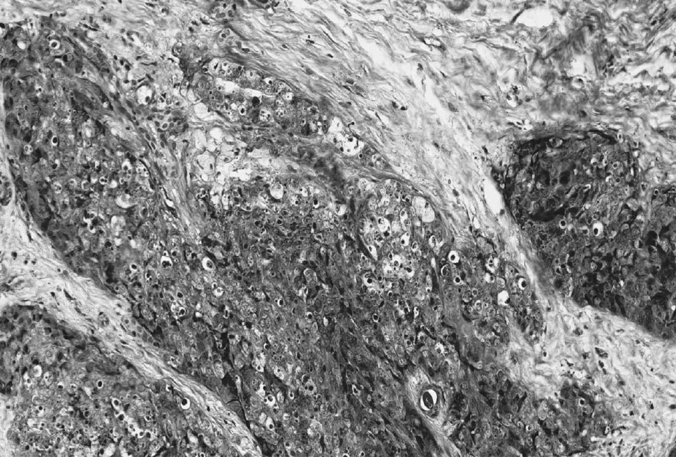
Figures reproduced from Wang J, et al. 2023, covered by a CC-BY-4.0 license: http://creativecommons.org/licenses/by/4.0/


Incidence and risk factors
When defining EMD as only involving soft-tissue plasmacytomas unrelated to bone1,4:
0.5%–4.8%
Incidence of MM with EMD at initial diagnosis*


3.4%–14%
Incidence of MM with EMD at relapse
In patients with MM, risk of EMD is higher with5:
- Osteolytic lesions
- Hypercalcemia
- Exposure to more lines of therapy
Incidence of EMD is higher in patients with5,6:
- High-risk cytogenetics
- High serum lactate dehydrogenase, indicative of highly proliferative disease
Incidence of EMD increases as5:
- Disease becomes more refractory
Location
While EMD can be found anywhere, in a study of 127 patients with MM and EMD relapse, the most common sites (≥10%) were7†:


MM with CNS involvement
- CNS EMD is a rare form of EMD characterized by plasma cell infiltration of the CNS, meninges or cerebrospinal fluid8
- Median OS for patients with EMD and CNS involvement ranges from 3-7.4 months9
*The reported incidence of EMD varies considerably, and differences in diagnostic approach between studies are likely to contribute to this variability. †For genitourinary organs, spleen and gums (n=1, 0.8% each), pancreas (n=3, 2%), gastrointestinal tract and breast (n=10, 8% each). ‡Always in addition to another site.
Diagnosis and treatment
Diagnosis

EMD can be detected by CT, PET, and MRI scans10§

CNS EMD can be detected by brain MRI and lumbar puncture8
Treatment
- Patients with EMD have high unmet medical need; MM-focused therapies include chemotherapy, radiotherapy, stem cell transplant, immunomodulatory agents, proteasome inhibitors, and MM antigen-targeting immunotherapies such as CAR-T, monoclonal antibodies, and bispecific antibodies1,2,11-13
- Treatment options for EMD with CNS involvement are limited. Systemic therapy, intrathecal therapy and cranial irradiation can be used, often in combination10
- Clinical trials dedicated to patients with EMD are exploring novel treatment regimens14
§When available,18fluorodeoxyglucose (18F-FDG) PET/CT is recommended by the International Myeloma Working Group for the workup of patients with newly diagnosed and relapsed MM.15 Use of PET/CT and MRI provides the highest probability of EMD detection by combining the anatomical and functional imaging capability of PET/CT with the high resolution of soft tissues and CNS structures from MRI.16
CAR-T, chimeric antigen receptor‑T cell; CELMod, Cereblon E3 ligase modulatory drugs; CNS, central nervous system; CT, computed tomography; EMD, extramedullary disease; EM-B, extramedullary-bone-related; EM-E, extramedullary-extraosseous; MM, multiple myeloma; MRI, magnetic resonance imaging; NDMM, newly diagnosed multiple myeloma; OS, overall survival; PET, positron emission tomography; PFS, progression-free survival.
1. Blade J, et al. Blood Cancer J. 2022;12(3):45. 2. Wang J, et al. Ann Med. 2023;55(2):2281657. 3. Holler A, et al. Cancer Med. 2022;11(24):4743–4755. 4. Beksac M, et al. Haematologica. 2020;105(1):201. 5. Bansal R, et al. Blood Cancer J. 2021;11(9):161. 6. Chawla Y, et al. Cancer Metab. 2025;13(1):9. 7. Avivi I, et al. Am J Hematol. 2019;94:1132–1140. 8. Egan PA, et al. Haematologica. 2020;105(7):1780–1790. 9. Tyczyńska A, et al. Biomedicines. 2023;11(4):1225. 10. Touzeau C, et al. Blood. 2016;127(8):971–976. 11. McAvera R, et al. Int J Mol Sci. 2023;24(14):11259. 12. Guliyev M, et al. Int J Hematol. 2025;122(1):73-82. 13. Sanchez L, et al. Semin Hematol. 2025;62(1):31- 37. 14. ClinicalTrials.gov. Accessed February 20, 2026. NCT06793475, NCT04586426, NCT04649359, NCT06518551, NCT06627751. 15. Cavo M, et al. Lancet Oncol. 2017;18(4):e206–2017. 16. Sevcikova S, et al. Blood Rev. 2019;36:32–39.
Connect with us
Connect with us
Would you like to clear and leave your conversation? Message history will be lost.